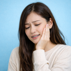
Schiffman E, Ohrbach R, Truelove E, et al. Diagnostic Criteria for Temporomandibular Disorders (DC/TMD) for Clinical and Research Applications. J Oral Facial Pain Headache. 2014;28(1):6-27.
Slade GD, Fillingim RB, Sanders AE, et al. Summary of findings from the OPPERA prospective cohort study of incidence of first-onset temporomandibular disorder. J Pain. 2013;14(12 Suppl):T116-T124.
Ooi K, Kashiwagi M, et al. Clinical practice guidelines in primary treatment for temporomandibular disorders: The Japanese Society for the Temporomandibular Joint, 2023 edition. J Prosthodont Res. 2025;69(4):608-617.
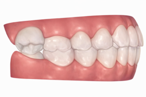
Manfredini D, Lombardo L, Siciliani G. Temporomandibular disorders and dental occlusion. A systematic review of association studies: end of an era? J Oral Rehabil. 2017;44(11):908-923.
Kim MR, Graber TM, Viana MA. Orthodontics and temporomandibular disorder: a meta-analysis. Am J Orthod Dentofacial Orthop. 2002;121(5):438-446.
Fernández-González FJ, Cañigral A, López-Caballo JL, et al. Influence of orthodontic treatment on temporomandibular disorders: a systematic review. J Clin Exp Dent. 2015;7(2):e320-e327.
Shalish M, Leibovich A, Zakuto A, et al. The association between orthodontic treatment and temporomandibular disorders diagnosis and disease characteristics. J Oral Rehabil. 2024;51(3):487-499.
Coronel-Zubiate FT, Marroquín-Soto C, Geraldo-Campos LA, et al. Association between orthodontic treatment and the occurrence of temporomandibular disorders: a systematic review and meta-analysis. J Clin Exp Dent. 2022;14(12):e1032-e1043.
Al-Moraissi EA, Farea R, Qasem KA, et al. Effectiveness of occlusal splint therapy in the management of temporomandibular disorders: network meta-analysis of randomized controlled trials. Int J Oral Maxillofac Surg. 2020;49(8):1042-1056.
Ebrahim S, Montoya L, Busse JW, et al. The effectiveness of splint therapy in patients with temporomandibular disorders: a systematic review and meta-analysis. J Am Dent Assoc. 2012;143(8):847-857.
Kulkarni S, Baig MS, Ansari Z, et al. Evaluating the effectiveness of nonsteroidal anti-inflammatory drug(s) for relief of pain associated with temporomandibular joint disorders: A systematic review. Clin Exp Dent Res. 2020;6(1):134-146.
Jiménez-Silva A, Peña-Durán C, Tobar-Reyes J, Frugone-Zambra R. Sleep and awake bruxism in adults and its relationship with temporomandibular disorders: a systematic review from 2003 to 2014. Acta Odontol Scand. 2017;75(1):36-58.
Mortazavi N, et al. Is bruxism associated with temporomandibular joint disorders? A systematic review and meta-analysis. Evid Based Dent. 2023;24(3):144.
Minervini G, et al. Prevalence of temporomandibular disorders in children and adolescents evaluated with Diagnostic Criteria for Temporomandibular Disorders: a systematic review with meta-analysis. J Oral Rehabil. 2023;50(6):522-530.
Poluha RL, De la Torre Canales G, Costa YM, et al. Temporomandibular joint disc displacement with reduction: a review of mechanisms and clinical presentation. J Appl Oral Sci. 2019;27:e20180433.
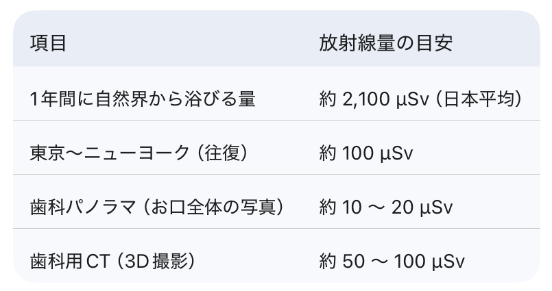
International Commission on Radiological Protection. The 2007 Recommendations of the International Commission on Radiological Protection (ICRP Publication 103). Ann ICRP. 2007.
Horner K, et al. Cone Beam CT for Dental and Maxillofacial Radiology: Evidence Based Guidelines (Radiation Protection No. 172). European Commission; 2012.
Pauwels R, et al. Effective dose range for dental cone beam computed tomography scanners. Dentomaxillofac Radiol. 2012.
Ludlow JB, Walker C. Effective dose of dental CBCT―A meta analysis of published data and additional data for nine CBCT units. Dentomaxillofac Radiol. 2014.
Shin HS, et al. Effective doses from panoramic radiography and CBCT using dose-area product. Imaging Sci Dent. 2014.
Grünheid T, et al. Dosimetry of a cone-beam computed tomography machine compared with conventional radiography. Am J Orthod Dentofacial Orthop. 2012.
Brooks SL. CBCT Dosimetry: Orthodontic Considerations. Oral Maxillofac Surg Clin North Am. 2009.
Pauwels R, et al. Cone beam CT for dental and maxillofacial imaging: dose matters. Radiat Prot Dosimetry. 2015.
Li G. Patient radiation dose and protection from cone-beam computed tomography. Imaging Sci Dent. 2013.
International Atomic Energy Agency. Radiation doses in dental radiology (RPOP). IAEA.
United Nations Scientific Committee on the Effects of Atomic Radiation. UNSCEAR 2024 Report, Volume I. 2024.
UK Government. Patient dose information: guidance (typical effective doses for diagnostic imaging). 2008.
American Dental Association. Updated recommendations to enhance radiography safety in dentistry. 2024.
American Academy of Oral and Maxillofacial Radiology. General Dental Imaging: Patient Shielding (brochure/position). 2024.
Benavides E, et al. Patient shielding during dentomaxillofacial radiography. Oral Surg Oral Med Oral Pathol Oral Radiol. 2023.
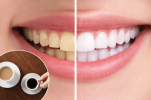
Watts A, Addy M. Tooth discolouration and staining: a review of the literature. Br Dent J. 2001;190(6):309-316. doi:10.1038/sj.bdj.4800959.
Sulieman M. An overview of tooth discoloration: extrinsic, intrinsic and internalized stains. Dent Update. 2005;32(8):463-471. doi:10.12968/denu.2005.32.8.463.
Carpenter GH, Pramanik R, Proctor GB. An in vitro model of chlorhexidine-induced tooth staining. J Periodontal Res. 2005;40(3):225-230. doi:10.1111/j.1600-0765.2005.00791.
Sarembe S, et al. Staining potential of common beverages using an in vitro staining and brushing model. Eur J Dent. 2022.
Wang J, et al. Drug-induced tooth discoloration: An analysis of the US FDA adverse event reporting system. Front Pharmacol. 2023.
Kumar A, et al. Drug-induced discoloration of teeth: an updated review. Clin Pediatr (Phila). 2012.
Enax J, et al. Toothpaste abrasion and abrasive particle content: correlating profilometry with RDA. Dent Mater J. 2023.
Vural UK, et al. Effects of charcoal-based whitening toothpastes on human enamel: color, roughness, microhardness (in vitro). Clin Oral Investig. 2021.
Osmanaj F, et al. Abrasion behavior of different charcoal toothpastes on human dentin when using electric toothbrushes. Clin Oral Investig. 2022.
Hongsathavij R, et al. Clinical comparison of the stain removal efficacy of two air polishing powders. Clin Cosmet Investig Dent. 2017.
Sigwart L, et al. Colour changes and surface roughness after air-polishing for tobacco stain removal. Int Dent J. 2025.
Talic NF, Almudhi AA. The effect of dietary pigmentation on the esthetic appearance of clear orthodontic elastomeric modules. J Orthod Sci. 2016;5(2).
da Silva VD, et al. Influence of food colorings on esthetic orthodontic elastomeric ligatures. J Appl Oral Sci. 2016.
Mazhari M, et al. Color stability of orthodontic elastomeric ligatures in the oral environment: a clinical trial. Front Dent. 2025.
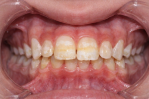
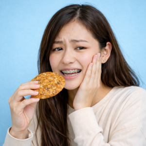
Khoroushi M, Kachuie M. Prevention and treatment of white spot lesions in orthodontic patients. Contemp Clin Dent. 2017;8(1):11-19.
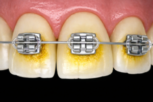
Cerroni S, Pasquantonio G, Condò R, Cerroni L. Orthodontic fixed appliance and periodontal status: an updated systematic review. Open Dent J. 2018;12:614.
Bollen AM, Cunha-Cruz J, Bakko DW, Huang GJ, Hujoel PP. The effects of orthodontic therapy on periodontal health: a systematic review of controlled evidence. J Am Dent Assoc. 2008;139(4):413-422.
Ristic M, Vlahovic Svabic M, Sasic M, Zelic O. Clinical and microbiological effects of fixed orthodontic appliances on periodontal tissues in adolescents. Orthod Craniofac Res. 2007;10(4):187-195.
Davies TM, Shaw WC, Worthington HV, Addy M, Dummer P, Kingdon A. The effect of orthodontic treatment on plaque and gingivitis. Am J Orthod Dentofacial Orthop. 1991;99(2):155-161.
Sukontapatipark W, el-Agroudi MA, Selliseth NJ, Thunold K, Selvig KA. Bacterial colonization associated with fixed orthodontic appliances: a scanning electron microscopy study. Eur J Orthod. 2001;23(5):475-484.
van Gastel J, Quirynen M, Teughels W, Coucke W, Carels C. Longitudinal changes in microbiology and clinical periodontal variables after placement of fixed orthodontic appliances. J Periodontol. 2008;79(11):2078-2086.
Enaia M, Bock N, Ruf S. White-spot lesions during multibracket appliance treatment: a challenge for clinical excellence. Am J Orthod Dentofacial Orthop. 2011;140(1):e17-e24.
Hussain U, Wahab A, Kamran MA, et al. Prevalence, incidence and risk factors of white spot lesions associated with orthodontic treatment: a systematic review and meta-analysis. Orthod Craniofac Res. 2025;28(2):379-399.
Sonesson M, Twetman S. Prevention of white spot lesions with fluoride varnish during orthodontic treatment with fixed appliances: a systematic review. Eur J Orthod. 2023;45(5):485-490.
Benson PE, Parkin N, Dyer F, Millett DT, Furness S, Germain P. Fluorides for the prevention of early tooth decay (demineralised white lesions) during fixed brace treatment. Cochrane Database Syst Rev. 2019;12.
Pithon MM, Baião FS, Sant’Anna LID, Tanaka OM, Cople-Maia L. Effectiveness of casein phosphopeptide-amorphous calcium phosphate-containing products in the prevention and treatment of white spot lesions in orthodontic patients: a systematic review. J Investig Clin Dent. 2019;10(2):e12391.
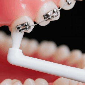
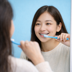
AlMoharib HS, Alqasem A, Almusfer G, Aldosari MA, Almadhoon HW. The effectiveness of water jet flossing and interdental flossing for oral hygiene in orthodontic patients with fixed appliances: a randomized clinical trial. BMC Oral Health. 2024;24(1):498.
Haeger RS, Colberg RT. Effects of missed appointments and bracket failures on treatment efficiency and office productivity. J Clin Orthod. 2007;41(8):433-437.
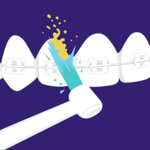
Huang J, Yao Y, Jiang J, Li C. Effects of motivational methods on oral hygiene of orthodontic patients: a systematic review and meta-analysis. Medicine (Baltimore). 2018;97(47):e13182.
Klukowska M, Bader A, Erbe C, et al. Plaque levels of patients with fixed orthodontic appliances measured by digital plaque image analysis. Am J Orthod Dentofacial Orthop. 2011;139(5):e463-e470.
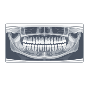
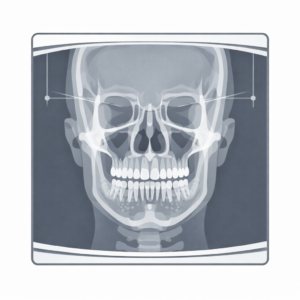
American Academy of Oral and Maxillofacial Radiology. Clinical recommendations regarding use of cone beam computed tomography in orthodontics. Oral Surg Oral Med Oral Pathol Oral Radiol. 2013;116(2):238-257.
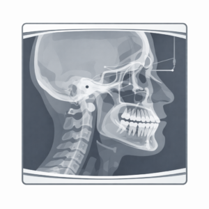
Kapetanović A, Theodorou C, Nilsson J, et al. Orthodontic radiology: development of a clinical practice guideline. Eur J Orthod. 2020;42(6):595-604.
Isaacson KG, Horner K, Whaites E. Guidelines for the Use of Radiographs in Clinical Orthodontics (4th ed.). British Orthodontic Society; 2016.
Ludlow JB, Timothy R, Walker C, et al. Effective dose of dental CBCT-a meta analysis of published data and additional data for nine CBCT units. Dentomaxillofac Radiol. 2015;44(1):20140197.
Suomalainen A, Kiljunen T, Käser Y, et al. Effective doses from panoramic radiography and cone beam computed tomography. Dentomaxillofac Radiol. 2014.
American Academy of Pediatric Dentistry. Prescribing dental radiographs for infants, children, adolescents, and individuals with special health care needs. The Reference Manual of Pediatric Dentistry. 2024/2025 update.
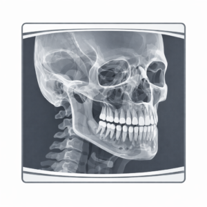
Eslami E, Barkhordar H, Abramovitch K, Kim J, Masoud MI. Cone-beam computed tomography vs conventional radiography in visualization of maxillary impacted-canine localization: A systematic review of comparative studies. Am J Orthod Dentofacial Orthop. 2017;151(2):248-258.
Guerrero ME, Shahbazian M, Elsiena Bekkering G, Nackaerts O, Jacobs R, Horner K. The diagnostic efficacy of cone beam CT for impacted teeth and associated features: a systematic review. J Oral Rehabil. 2011;38(3):208-216.
Haney E, Gansky SA, Lee JS, et al. Comparative analysis of traditional radiographs and cone-beam computed tomography volumetric images in the diagnosis and treatment planning of maxillary impacted canines. Am J Orthod Dentofacial Orthop. 2010;137(5):590-597.
Edmonds M, et al. Ability of orthodontists to detect, interpret and propose management strategies for incidental findings on pre-treatment panoramic radiographs. Orthod Craniofac Res. 2025.
Hlongwa P, Moshaoa MAL, Musemwa C, Khammissa RAG. Incidental pathologic findings from orthodontic pretreatment panoramic radiographs. Int J Environ Res Public Health. 2023;20(4):3479.
Leung CC, Palomo L, Griffith R, Hans MG. Accuracy and reliability of cone-beam computed tomography for measuring alveolar bone height and detecting bony dehiscences and fenestrations. Am J Orthod Dentofacial Orthop. 2010;137(4 Suppl):S109-S119.
Molen AD. Considerations in the use of cone-beam computed tomography for buccal bone measurements. Am J Orthod Dentofacial Orthop. 2010;137(Suppl):S130-S135.
Kapila SD, Nervina JM. CBCT in orthodontics: assessment of treatment outcomes and indications for its use. Dentomaxillofac Radiol. 2015.
European Journal of Orthodontics. Development of a clinical practice guideline for orthodontically induced external apical root resorption. 2019;42(2):115-123.
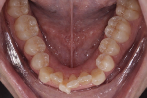
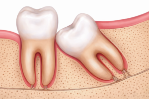
Lyros I, et al. The Effect of Third Molars on the Mandibular Anterior Crowding Relapse after Orthodontic Treatment: A Systematic Review. Dent J (Basel). 2023;11(5):131.
Harradine NW, Pearson MH, Toth B. The effect of extraction of third molars on late lower incisor crowding: a randomized controlled trial. Br J Orthod. 1998;25(2):117-122.
Žigante M, Pavlić A, Morelato L, Vandevska-Radunovic V, Spalj S. Presence and Maturation Dynamics of Mandibular Third Molars and Their Influence on Late Mandibular Incisor Crowding: A Longitudinal Study. Int J Environ Res Public Health. 2021;18(19):10070.
Cheng HC, Peng BY, Tam KW. Impact of third molars on mandibular relapse in post-orthodontic patients: A meta-analysis. J Dent Sci. 2018;13(1):1-7.
Little RM. The irregularity index: a quantitative score of mandibular anterior alignment. Am J Orthod. 1975;68(5):554-563.
Ghaeminia H, et al. Surgical removal versus retention for the management of asymptomatic disease-free impacted wisdom teeth. Cochrane Database Syst Rev. 2020;5:CD003879.
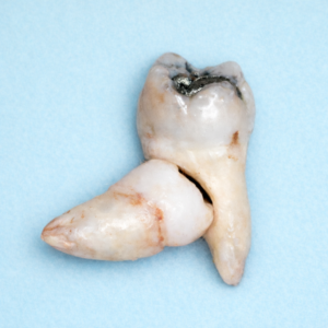
Revuelta-Cortés P, Cortés-Bretón Brinkmann J, Argandoña-Flores M, et al. Prevalence of distal caries in second molar associated with impacted mandibular third molar and the position and level of impaction: a systematic review and meta-analysis. Clin Oral Investig. 2025;29(1):83.
Ma Y, Mu D, Li X. Risk factors for root resorption of second molars with impacted third molars: a meta-analysis of CBCT studies. Acta Odontol Scand. 2023;81(1):18-28.
Oenning ACC, et al. External Root Resorption of the Second Molar Associated with Third Molar Impaction: A Comparison Using Cone-Beam Computed Tomography. J Oral Maxillofac Surg. 2014.
Sarikov R, Juodzbalys G. Inferior alveolar nerve injury after mandibular third molar extraction: a literature review. J Oral Maxillofac Res. 2014;5(4):e1.
American Association of Oral and Maxillofacial Surgeons. Management of Third Molar Teeth (White Paper). AAOMS; 2024.
National Institute for Health and Care Excellence (NICE). Guidance on the Extraction of Wisdom Teeth (TA1). NICE; 2000.
Miclotte A, Grommen B, Cadenas de Llano-Pérula M, et al. The effect of first and second premolar extractions on third molars: A retrospective longitudinal study. J Dent. 2017;61:55-66.
Zhu J, et al. Comparison of postoperative outcomes between envelope and triangular flaps after mandibular third molar surgery: A systematic review and meta-analysis. J Oral Maxillofac Surg. 2020.
Tsichlaki A, Chin SY, Pandis N, et al. How long does treatment with fixed orthodontic appliances take? A systematic review. Eur J Orthod. 2016;38(3):325-330.
Mavreas D, et al. Factors affecting the duration of orthodontic treatment: a systematic review. Eur J Orthod. 2008;30(4):386-395.
Fink D, Smith R. The duration of orthodontic treatment. Am J Orthod Dentofacial Orthop. 1992;102(5):391-396.
Abbing D, et al. Duration of orthodontic treatment with fixed appliances: a systematic review. Prog Orthod. 2020;21:27.
Wazwaz F, et al. Duration of tooth alignment with fixed appliances: a systematic review. Am J Orthod Dentofacial Orthop. 2022;161(5):669-679.
Beckwith FR, et al. An evaluation of factors affecting orthodontic treatment duration. Am J Orthod Dentofacial Orthop. 1999;116(1):17-26.
Moresca R. Orthodontic treatment time: can it be shortened? Prog Orthod. 2018;19:30.
Abbing D, et al. Treatment duration fixed appliances in adolescents vs adults: meta-analysis. Prog Orthod. 2020;21:30.
Minimal important difference in orthodontic treatment duration. Eur J Orthod. 2024.
Orthodontic treatment with clear aligners vs fixed appliances systematic review. J Orthod Sci. 2024.
Optimal treatment timing in orthodontics review. PMC. 2008.
Factors affecting orthodontic treatment time review (2023). IJCMPh. 2023.
Influence of orthodontist change on treatment duration. BMC Oral Health. 2024.
Orthodontic treatment in adults: challenges and outcomes. PMC. 2024.
American Dental Association. Pregnancy. Updated July 14, 2025. Accessed December 23, 2025.
American College of Obstetricians and Gynecologists. Oral Health Care During Pregnancy and Through the Lifespan. Committee Opinion No. 569. Obstet Gynecol. 2013;122(2 Pt 1):417-422.
American Academy of Pediatric Dentistry. Oral Health Care for the Pregnant Pediatric Dental Patient. The Reference Manual of Pediatric Dentistry. 2025.
Manautou MA, Mayberry ME. Local anesthetics and pregnancy: a review of pharmacology and clinical considerations. Anesth Prog. 2023.
Lopes IC, et al. Analgesia in pregnant patients: a literature review. 2024.
Zhang J, et al. Expert consensus on the treatment of oral diseases in pregnancy. 2025.
Mukherjee PM, Almas K. Orthodontic considerations for gingival health during pregnancy. J Clin Periodontol. 2010;37(8):696-702.
FDI World Dental Federation. Oral Health and Pregnancy: Fact Sheet. 2024/2025.
Massachusetts Department of Public Health. Oral Health Practice Guidelines for Pregnancy & Early Childhood. 2024.
ADA. Nitrous Oxide Safety for Pregnant Dental Staff and Patients. Accessed December 23, 2025.
Khinda V, et al. Nitrous oxide inhalation sedation: clinical guidance and safety considerations. 2023.
ACOG. Guidelines for Diagnostic Imaging During Pregnancy and Lactation (overview statements). Accessed December 23, 2025.
Silk H, Douglass AB, Douglass JM, Silk L. Oral health during pregnancy. Am Fam Physician. 2008;77(8):1139-1144.
Gajendra S, Kumar JV. Oral health and pregnancy: a review. N Y State Dent J. 2004;70(1):40-44.
Offenbacher S, et al. Periodontal disease and adverse pregnancy outcomes: epidemiology and biological plausibility. Periodontol 2000. 2013;62(1):125-139.